Showing posts with label tregs. Show all posts

Thursday, March 15, 2012

Redirecting the Melanoma Tumor’s Microenvironment in Favor of the Activation of T-cells..Jim Breitfeller


Studies show that regulatory T (Treg) cells play a detrimental role in cancer immunotherapy because these cells accumulate in the tumor microenvironment and suppress immune responses. Moreover, Researchers recently showed the presence of tumor-specific CD4+, CD8+, and γδ Treg cells in several types of tumors, suggesting that they can induce antigen-specific, local immune tolerance at tumor sites. Tumor-associated macrophages (TAMs) and myeloid-derived suppressor cells (MSC) could also play an important role in inhibiting immune responses and chronic inflammation, which has been linked to cancer development and progression. Both tumor-associated macrophages/DCs and MSCs promote tumor growth either by secreting immunosuppressive cytokines, including interleukin 10 (IL-10), transforming growth factor-β (TGF-β), and IL-1β, or by inducing Treg cell differentiation. More importantly, tumor cells have been shown to express inhibitory factors (IL-10, TGF-β, GAL-3, and IDO) to alter T-cell function. Immunosuppressive factors, such as FasL and TGF-β expressed by tumor cells, may directly inhibit tumor-reactive T-cell expansion or induce T-cell apoptosis. A recent studies suggest that tumor-associated galectin-1(Gal-1) and or Gal-3, a membes of the animal lectin family, contributes to tumor immune escapes by inhibiting the function of tumor-reactive T cells. Therefore, tumor cells constantly modulate T-cell responses by presenting tumor antigens and secreting immunoregulatory cytokines. Understanding the interplay between tumor cells and immune cells in the tumor microenvironment is essential for the development of effective cancer immunotherapy.

Researchers tested whether Gal-3 could activate other tumor-reactive or antigen-experienced T cells. Five melanoma-reactive T-cell lines, one prostate cancer–derived T-cell line, and two breast cancer–derived T-cell lines were selected and cocultured with 293 cells expressing Gal-3 for 12 to 16 h. they found that Gal-3–expressing 293 cells activated all of these T-cell lines to secrete IFN-γ but failed to activate naive CD4+ and CD8+ T cells purified from peripheral blood mononuclear cells (PBMC) of healthy donors . Suggesting that naive T-cell activation requires a strong T-cell receptor (TCR)-mediated activation, whereas tumor-reactive T cells can be readily activated by Gal-3. They also evaluated the cytokine profiles of CT28 T cells on Gal-3 stimulation. Gal-3 induced a high level of IFN-γ, granulocyte macrophage colony-stimulating factor, and low to middle levels of IL-4, which is similar to cytokine production induced by anti-CD3 (OKT3) stimulation. This is an indication that the Gal-3 complexes with the TCR synapse causing impaired signaling. Gal-3 binds and activates tumor-reactive T cells through carbohydrate-specific interaction.

Galectin-3 (GAL-3) localize mainly in Tumor cells, macrophages, epithelial cells,
Fibroblasts , and activated T-cells. Although galectin-1 has been shown to induce T-cell apoptosis, galectin-3 has conversely been shown to prevent cell death induced by Fas ligation. Galectin-3 has been shown to rescue cells from apoptosis by protecting against alterations of the mitochondrial membrane and formation of reactive oxygen species. A growing body of evidence supports the involvement of galectin-3 in tumor growth and metastasis.

Galectin-3 and IL-10 receptor needs to be inhibited to break the rest of the tolerance so the immunotherapy can have a greater effect with a better response rate.

Radiation + Yervoy + Anti-PD-1 + Anti-IL-10 receptor + GAL-3 Inhibitor= Robust immune response


“It is not the strongest of the species that survives, nor the most intelligent, but the one most responsive to change.”

~Charles Darwin~
Take Care,
Jimmy B
Photobucket

Friday, August 26, 2011

How does Ipilimumab (Yervoy) work? Melanoma..Jim Breitfeller

Ipilimumab is anti-CTLA-4 monoclonal antibody.

Ipilimumab C6742H9972N1732O2004S40
immunoglobul has a calculated molecular formula of C6472H9972N1732O2004S40 and a molecular weight of 145.4 kDa.in G1, anti-(human CTLA-4 (antigen)) (human gamma1-chain), disulfide with human kappa-chain, dimer [CAS] immunoglobulin G1, anti-(human CTLA-4 (antigen)) (human g1-chain), disulfide with human k-chain, dimer [CAS]

Half Life = 15 days

Tremelimumab (from Pfizer) is an IgG2
Tremelimumab is a fully human cytotoxic T lymphocyte-associated antigen-4 (CTLA-4) IgG2 monoclonal antibody, presumably manufactured using transformed mammalian cells. Tremelimumab is a dimer (composed of two chains) linked by a disulfide/sulfhydryl bond. Tremelimumab has a calculated molecular formula of C6500H9974N1726O2026S52 and a molecular weight of ~150 kDa

Half life= 21 days


In T-cell activation, the receptor, CTLA-4 is upregulated on about day 3.

The CTLA-4 complex can suppress the immune response.

CTLA-4 that interferes with Dendritic cell (DC) costimulation via reduced CD80/CD86, (B7-1-2) expression and CD25 (IL-2 receptor) to allow for Treg survival, activation, and effective competition for limited IL-2 during infection.

Ipilimumab (Yervoy) works by blocking the CTLA-4/ B7-1/2 complex, which allows the costimulation of the CD28// B7-1/2 complex to take place. This takes the brakes off the immune response allow the response to go unchecked.

Another thing Yervoy does is that it decreases the signaling of STAT5 with increasing concentrations. That is why the best overall concentration is 10mg/Kg.

Phosphorylation of STAT5 decreased significantly with increasing concentrations of tremelimumab/Ipilimumab in monocytes from five healthy donors and from three patients with melanoma. Overall, these data demonstrate that monocytes express mostly intracellular CTLA4 and that CTLA4 is biologically active in this cell subset since exposure to tremelimumab/ipilimumab induces changes in intracellular signaling molecules.
By decreasing the signaling of STAT5 during Cell differentiation, TH17 and TH1 cells are mostly produced.


“It is not the strongest of the species that survives, nor the most intelligent, but the one most responsive to change.”
~Charles Darwin~
Take Care,
Jimmy B

Photobucket

Friday, October 1, 2010

Tregs Rule Unless Anti-CTLA-4 Blockage is Used!!! Melanoma.. Jim Breitfeller


The first step is to relieve the immune system from the tumor-dependent immune tolerance mediated by Treg, which prevents the development of an efficient antitumor immune response.

The second step is the activation of the immune response.


“It is not the strongest of the species that survives,
nor the most intelligent, but the one most responsive to change.”


~Charles Darwin~

Take Care,

Jimmy B

Photobucket

Friday, September 24, 2010

Living Medical Textbook: Update to the Tumor Immunology and Immunotherapy: Metastatic Melanoma ..Jim Breitfeller

Living Medical Textbook: Update to the Tumor Immunology and Immunotherapy: Metastatic Melanoma Edition With Dr. Jeffery Weber and Dr. Bernard Fox

Jeffrey S. Weber, MD, PhD
Senior Member, Moffitt Cancer Center
Director, Comprehensive Melanoma Research Center
Professor, Department of Oncologic Sciences
H. Lee Moffitt Cancer Center & Research Institute
Tampa, Florida


Bernard A. Fox, PhD
Chief
Laboratory of Molecular and Tumor Immunology
Providence Portland Medical Center
Earle A. Chiles Research Institute
Portland, Oregon


This "living" textbook is an interactive and up-to-date resource to support learning in the field of tumor immunology, and more specifically focuses on melanoma treatment and management. Throughout each chapter you will find supplemental hyperlinks to external multimedia resources—including illustrations, ...more


Source:http://www.livingmedicaltextbook.org/Activity/index.cfm?jn=2001&sj=2001.01&i=5

You must turn off my playlist by clicking on the radio button on my playlist located on the right column almost all the way down the scrollbar

Listen as Dr. Weber interviews Dr. Fox about the ability of helper T cells, cytolytic T cells, and natural killer T cells to mediate tumor regression.


Listen as Dr. Weber interviews Dr. Fox about the ability of regulatory T cells to dampen the immune response to tumor cells.


Listen as Dr. Weber and Dr. Fox discuss investigational immunotherapies that improve T cell targeting of tumors.


Special Feature: Replay the complete audio interview between Dr. Weber and Dr. Fox


“It is not the strongest of the species that survives, nor the most intelligent, but the one most responsive to change.”
~Charles Darwin~

Take Care,

Jimmy B

Photobucket

Tuesday, September 21, 2010

The missing third signal "The Danger Signal"Melanoma..Jim Breitfeller

Th17 Cells Secrete IL-17 in the Tumor Microenvironment causing inflammatory symptoms "The Danger Signal"improves Survival in a Murine Model of Pancreatic Cancer and in Melanoma also.

Third, inflammatory cytokines, including IL-1, IL-6, IL-12, and IFN-γ provide a third signal that acts directly on T cells, referred to as the “danger signal”. This signal was found to optimally activate TH1 differentiation and lead to clonal expansion of T cells.

"Th17 cells and IL-17 participate in antitumor immunity by facilitating T cell recruitment to the tumor site and CD8+ T cell priming and effector differentiation suggests a new avenue for developing Th17 cell-based therapy."

~S. A. Rosenberg~


"Using in vitro and in vivo approaches, we determined that under neutral conditions, simultaneous activation of Tregs and naive CD4+ conventional T cells in the presence of APCs resulted in conversion of Tregs into IL-17–producing cells, and endogenous IL-1β was mandatory in this process" according to Vassiliki A. Boussiotis et al


Thus, the addition of IL-6 and IL-1β to the tumor microenvironment skews the balance toward Th17 cells in a murine model of pancreatic cancer and Melanoma.

So we need to suppress the Tregs and generate Th17 T-cells to initiate the right immune response.


“It is not the strongest of the species that survives, nor the most intelligent, but the one most responsive to change.”
~Charles Darwin~
Take Care,
Jimmy B
Photobucket

Sunday, September 19, 2010

The Missing Link in T-cell activation using a Vaccine, "The Danger Signal"Melanoma..Jim Breitfeller

As I research why some patients respond to therapies i.e. vaccination and other immunotherapy and others don’t, I ask WHY? In my quest to get the answer or answers, I came across a paper called “Marked Differences in Human Melanoma Antigen-Specific T Cell Responsiveness after Vaccination Using a Functional Microarray”.

Daniel S. Chen1,2#, Yoav Soen3#, Tor B. Stuge4, Peter P. Lee4, Jeffrey S. Weber5, Patrick O. Brown2,3, Mark M. Davis2,6*

1 Department of Internal Medicine/Division of Oncology, Stanford University, Stanford, California, United States of America, 2 Howard Hughes Medical Institute, Stanford University, Stanford, California, United States of America, 3 Department of Biochemistry, Stanford University, Stanford, California, United States of America, 4 Department of Medicine, Stanford University, Stanford, California, United States of America, 5 Norris Cancer Center, University of Southern California, Los Angeles, California, United States of America, 6 Department of Microbiology and Immunology, Stanford University, Stanford, California, United States of America


This is what I was looking for. It may hold the answer or could possibly point me in the right direction.

In the paper I came across a diagram that peaked my interest. It was a comparison between responders and non-responders.


They were looking at the cytokines secreted after the vaccine was given. When I saw what the cytokines were, I knew I was on the right track. These cytokines help in the differentiation of the CD4+ T-cells. What a find!!


Naïve CD4 T cells in the presence of TGF-b and IL-2 and others differentiate into Tregs.

TGF-b accelerates the CTLA-4 expression by stimulated CD4+ CD25- T-cells.


TGF-b requires CTLA-4 early after T-cell activation to induce FoxP expression generating CD4+ CD25+ Treg Regulatory cells.

The Th-17 cells produce IL-17. .IL-17 induces the production of many other cytokines (such as IL-6, G-CSF, GM-CSF, IL-1β, TGF-β, TNF-α).


So what was the non-responder missing, IL-6. With the missing IL-6, they weren’t able to produce Th-17 that secreted IL-17.


While TGF-β is a critical differentiation factor for Treg cells, IL6 completely inhibits the generation of Treg cells induced by TGF-β. Instead, IL6 and TGF-β together induce the differentiation of pathogenic Th17 cells. With IL-6 missing in the microenvironment, Treg Cells flourish.

If the CD4 + T cells differentiate into TH2 cells that produce IL-4, the other cells inhibited to produce IL-6. IL-4 was found to inhibit TNF-α and IL-1β by activated monocytes almost 100 %. The Secretion of IL-6 was decreased by approximatly 80 % in the presences of IL-4 Cytokine. TE Velde et al 1990

They were missing “The Danger Signal”.


Friendly inflammation “The Danger Signal”

Most of the time you have no notion of the microbial life-and-death struggle being waged within your body. At other times, though, you are acutely aware of the exact location of the battleground, thanks to the unmistakable signs of inflammation — heat, pain, redness, and swelling. Inflammation, the buildup of fluid and cells at the point of infection/cancer, is put into motion by cytokines — proteins that are released into the blood by the innate immune system when it encounters germs. Cytokines function like police dispatchers. They signal there's a problem, which activates the immune system's highway patrol force: the circulating lymphocytes of the adaptive immune system. These lymphocytes cruise the highways of the blood vessels and lymphatic system. In response to the chemical signal from the cytokines, increased blood flow rushes these circulating cells to the trouble spot.

“The CD8+ T-cell-mediated Immune Response to Eradicate the Tumors”

“Three major events must occur to induce CD8+ T cell–mediated, tumor-protective immunity against syngeneic melanoma. First, the T-cell receptor must be triggered by a (or multiple) self antigen–derived peptide MHC class I complex . Therefore, this event depends entirely on appropriate antigen presentation, which is most efficiently provided by mature dendritic cells. Peripherally tolerant or “ignorant” self-reactive T-cell clones, once properly activated, may serve as tumor-specific effector T cells .Second, simultaneously with T-cell receptor triggering, a distinct second costimulatory signal must be delivered, mediated by IL-2, B7-1, or B7-2, which engage IL-2 receptors and CD28 on the surface of the T cell, respectively. A source of these cofactors for effective CD8+ T-cell stimulation can be provided by CD4+ T cells that release critical amounts of IL-2, or by mature dendritic cells that display an increased level of B7-1/B7-2 costimulatory molecules on their cell surfaces. Third, inflammatory cytokines, including IL-1, IL-6, IL-12, IL-17 and IFN-γ provide a third signal that acts directly on T cells, referred to as the “danger signal”.

This signal was found to optimally activate TH1 differentiation and lead to clonal expansion of T cells.


The responder was able to produce inflammatory cytokines, including IL-1, IL-6, IL-12, IL-17 and IFN-γ provide a third signal that acts directly on T cells, referred to as the “danger signal”.


This signal was found to optimally activate TH1 differentiation and lead to clonal expansion of T cells and invoke a robust immune response to the Melanoma Cancer.

Our data altogether suggest the following model: Th17 cells go to the tumor site and, by secreting IL-17, activates residential cells to produce CCL2 and CCL20, which provokes the mobilization of DCs and other leukocytes to the tumor site. DCs uptake tumor antigens in the lung or tumor site and migrate to the lymph nodes where they activate CD8+ T cells against the tumor. The new wave of effector CD8+ T cells migrates back to the lung or tumor site and kills established tumors.

Therefore, our data demonstrating that Th17 cells and IL17 participate in antitumor immunity by facilitating T cell recruitment to the tumor site and CD8+ T cell priming and effector differentiation suggests a new avenue for developing Th17 cell-based therapy for tumors or chronic viral infections and as an adjuvant for vaccinations.”

(Dudley and Rosenberg, 2007; Rosenberg and Dudley, 2004)


Conclusion: We not only want to supress the Tregs, we also need to produce the "Danger Signal" to optimally activate TH1 differentiation and lead to clonal expansion of T cells to generate a robust tumor-specific immune response.


The Missing Link in T-cell activation using a Vaccine, "The Danger Signal"


“It is not the strongest of the species that survives, nor the most intelligent, but the one most responsive to change.”

~Charles Darwin~

Take Care,
Jimmy B

Photobucket

Saturday, September 18, 2010

Poised for progress..Melanoma ..Jim Breitfeller

Poised for progress
By Bill Schaller

Dana-Farber Cancer Institute

Friday, September 17, 2010

A new focus on the immune system’s ability to both unleash and restrain its attack on disease has led scientists at Harvard-affiliated Dana-Farber Cancer Institute to identify cells in mice that prevent the immune system from attacking the animals’ own cells, protecting them from autoimmune diseases such as multiple sclerosis, Type 1 diabetes, and lupus.

The discovery, recently reported by the journal Nature, may give scientists an effective way of operating the immune system’s internal “control panel,” leading to improved therapies for a variety of diseases — from vaccines that prompt the immune system to stage a sustained assault on cancers, to treatments that derail the biological onslaught associated with autoimmune diseases. The fact that human immune system cells share key features with those in mice makes the prospect of such advances quite realistic, the study authors say.

“The traditional view of the immune system is of specialized groups of cells poised to attack foreign pathogens [disease-causing agents],” said senior author Harvey Cantor, the Baruj Benacerraf Professor of Pathology at Harvard Medical School and chair of the Department of Cancer Immunology and AIDS at Dana-Farber. “While that model is generally correct, we’ve come to appreciate that the immune system, like other complex biological information systems, includes a counterbalance mechanism — a set of cells programmed to suppress the immune response. Such cells are essential to preventing excessive reactions to pathogens and misguided attacks on the body’s own cells.”

The search for cells involved in quieting the immune response has previously focused on immune system cells known as CD4+ T cells, some of which have been shown to prevent abnormal inflammation in response to disease or infection. In the new study, lead author Hye-Jung Kim and her colleagues found that CD8+ T cells (known as killer T cells because of their ability to kill diseased cells) also include a subset that helps dampen the immune response. Instead of reducing inflammation like their CD4 cousins, the CD8+ T regulatory (CD8+Treg) cells ensure that the immune system doesn’t produce antibodies that attack normal cells.

The Dana-Farber team discovered how CD8+ Treg cells accomplish this feat. They mingle with cells known as follicular T-helper cells, which are intermediaries that prompt the immune system’s B cells to make disease-fighting antibodies. The meeting with CD8+ Treg cells essentially shuts off the follicular T-helper cells, preventing them from interacting with B cells. No interaction means no production of antibodies, which means no assault on an animal’s normal, healthy cells.

The critical point of contact between CD8+ Treg cells and follicular T-helper cells is a protein on the helper cells called Qa-1. When Kim and her colleagues bred a strain of mouse with abnormal Qa-1, the animals developed a form of lupus. The reason: the CD8+ Treg cells couldn’t latch onto the defective protein, leaving the follicular cells free to order the B cells to produce antibodies, some of which targeted the animals’ own tissue.

The significance of this work is that CD8+ Treg cells represent a new lever for raising or lowering the strength of the immune response. This class of cells, it turns out, depends for its survival on a cytokine (a regulatory compound) called interleukin 15. Increase the supply of CD8+ Treg cells and the immune response is suppressed — a potentially powerful way of dealing with autoimmune diseases. Decrease the amount of such cells and the immune response can be invigorated and extended — a useful complement to vaccines that unleash the immune system on cancer.

“Experience has shown that vaccines that simply activate or expand the number of T and B cells are not likely to result in a prolonged, robust anti-tumor response,” Cantor explains. “The balancing mechanism within the immune system means that when more disease-fighting cells are generated, there’s a countervailing increase in the number of immune-suppressing cells that are generated. The key is to break that loop. This work brings that goal closer.”

Source:http://news.harvard.edu/gazette/story/2010/09/poised-for-progress/

A Phase I Study of Intravenous Recombinant Human IL-15 in Adults With Refractory Metastatic Malignant Melanoma and Metastatic Renal Cell Cancer
It is recruiting.

A Phase I Study of Intravenous Recombinant Human IL-15 in Adults With Refractory Metastatic Malignant Melanoma and Metastatic Renal Cell Cancer

Maybe a combination of Anti-CTLA-4 blockage + IL-15 may be another protocol that will erradicate the Melanoma Tumor


“It is not the strongest of the species that survives, nor the most intelligent, but the one most responsive to change.”

~Charles Darwin~

Take Care,
Jimmy B

Photobucket

Saturday, July 31, 2010

In Stage IV melanoma patients, a high percentage of Tregs appears to be associated with shorter survival. Jim Breitfeller

In Stage IV melanoma patients, a high percentage of Tregs appears to be associated with shorter survival.

Received 12 February 2008 published online 03 June 2008.

Background

Melanoma often elicits a profound immune response, and this response has been exploited by various immune therapies. These immunotherapies ultimately fail, however, and advanced melanoma is uniformly fatal, suggesting the development of an immune escape mechanism. In this study, markers of immune escape including regulatory T cells (Tregs), dendritic cells (DCs), and TGF-β were evaluated in 14 Stage IV melanoma patients and correlated with survival.

Source:http://www.journalofsurgicalresearch.com/article/S0022-4804(08)00344-2/abstract

In Stage IV melanoma patients, a high percentage of Tregs appears to be associated with shorter survival


Just follow the Science!!!!!!


Results

Stage IV melanoma patients had a doubling of regulatory T cells compared to both normal subjects and stage I melanoma patients. There was a significantly higher number of DCs in all melanoma patients compared to normal subjects. Stage I melanoma patients had a significantly higher number of pDCs than normal subjects, and all melanoma patients had a higher concentration of mDCs than controls. Serum IL-4 and IL-10 were not detectable but serum TGF-β levels were significantly higher in stage I and stage IV melanoma patients compared to normal controls.

Conclusion

Advanced melanoma is associated with increased numbers of circulating dendritic cells and regulatory T cells. These data suggest that melanoma induces immunosuppressive DCs and regulatory T cells in the systemic circulation.


Source:http://www.springerlink.com/content/k0gk8740u3n75744/


“It is not the strongest of the species that survives, nor the most intelligent, but the one most responsive to change.”
~Charles Darwin~

Take Care,
Jimmy B

Photobucket

Tregs and rethinking cancer immunotherapy..Melanoma..Jim Breitfeller

Tregs and rethinking cancer immunotherapy

Tyler J. Curiel

San Antonio Cancer Institute, University of Texas Health Sciences Center, and Cancer Therapy & Research Center, San Antonio, Texas, USA.
Address correspondence to: Tyler J. Curiel, San Antonio Cancer Institute, University of Texas Health Sciences Center, 7703 Floyd Curl Drive, San Antonio, Texas 78229-3900, USA. Phone: (210) 562-5286; Fax: (210) 562-5292; E-mail: curielt@uthscsa.edu.


Abstract
Introduction

Tumors express antigens that should induce immune-mediated rejection, but spontaneous rejection of established tumors is rare. Recent work demonstrates that one reason for the lack of tumor rejection is that tumors actively defeat host immunity. This concept forces us to rethink current approaches to harnessing potent, specific host immunity to battle cancer, most of which are based on the paradigm that inducing more antitumor immune cells alone is therapeutic. However, as I discuss in this Personal Perspective, a newer paradigm predicts that reducing tumor-driven immune suppression will be clinically beneficial. CD4+CD25+ Tregs are one mechanism of tumor-driven immune evasion that provide prototypical targets for testing novel anticancer treatment strategies within the newer paradigm.

Source:http://www.ncbi.nlm.nih.gov/pmc/articles/PMC1857250/

Tregs and rethinking cancer immunotherapyA>


This paper is one of the better ones for understanding the whole Treg Thing. Ipilimumab and Interluekin-2 in combination will change the paradigm in novel anticancer treatment strategies as we see it today and may hold the key to durable remissions of melanoma.


“It is not the strongest of the species that survives, nor the most intelligent, but the one most responsive to change.”

~Charles Darwin~


Take Care,
Jimmy B

Photobucket

Monday, July 26, 2010

Tamed T-Cells leave patients cancer-free..Melanoma..Jim Breitfeller

The science is coming in loud and clear. Remember I said blame it on the Tregs!!!!! Well Here is some conformation of that.

Tamed T-Cells leave patients cancer-free
Monday, 26 July 2010by Becky Crew


The antigen-specific reaction of T-cells working with dendritic cells, which regulate the immune system.


SYDNEY: The manipulation of specific immune cells in bone marrow transplants has left a number of patients cancer-free, giving new hope to leukemia and lymphoma sufferers, according to a recent study.

A team lead by Sébastien Maury from the Service d'Hématologie Clinique, France, has shown for the first time that by depleting the regulatory T-cell (or Treg) population in donor tissue, patients suffering from very resistant forms of cancer can achieve complete remission.

By identifying the Tregs’ influence on how a new immune system reacts to foreign elements, it’s hoped that this study will result in more successful cancer treatments using transplantation.

Source:http://www.cosmosmagazine.com/news/3586/tamed-t-cells-leave-patients-cancer-free
Tamed T-Cells leave patients cancer-free


This study is proof of a concept which demonstrates that regulatory T-cell manipulation can have therapeutic effects in humans,” says Maury. “It opens the way to its application in various fields in medicine where the immune system needs to be ‘tolerised’ (by adding more regulatory T-cells) in autoimmune diseases or ‘un-tolerised’ (by their elimination) in cancer.”


“It is not the strongest of the species that survives, nor the most intelligent, but the one most responsive to change.”
~Charles Darwin~

Take Care,
Jimmy B

Photobucket

Friday, May 21, 2010

High Dose Interluekin-2 followed by low-dose metronomic temozolomide (TMZ) to induce a complete immune Response.Melanoma ..Jim Breitfeller

High Dose Interluekin-2 followed by low-dose metronomic temozolomide (TMZ) to induce a complete immune Response

Background
CD4+CD25+ regulatory T cells (Treg), which constitute about 2–3% of CD4+ human T cells, are the main contributors to the maintenance of immune tolerance. Cancer patients, including Melanoma patients bear increased number of circulating and tumor infiltrating Treg that exert functional inhibition on tumor-specific T cells.

Temozolomide (sometimes referred to as TMZ) is an imidazotetrazine derivative of the alkylating agent dacarbazine.

Immunological factors relating to the antitumor effect of temozolomide ... appeared to suppress the frequency of CD4(+) CD25(+) regulatory T cells (Treg).
So base on the research, if activation occurs (mmune activation induced by the HD IL-2) IL-2 being a growth factor for the T-cells, will help fuel their expansion. Since the Tregs are a subset of the CD4+ T-cells, they too will proliferate.

By adding TMZ to the protocol, it suppresses the Treg function and shifts the balance tolerance toward activation of an immune response.

See the abstract at the ASCO Symposium 2010 "Ability of unsuccessful high-dose IL-2 therapy followed immediately by low-dose metronomic temozolomide to induce complete and near-complete remissions in metastatic melanoma."


Source:http://www.abstract.asco.org/AbstView_74_41534.html


“It is not the strongest of the species that survives, nor the most intelligent, but the one most responsive to change.”

~Charles Darwin~

Take Care,
Jimmy B
Photobucket

Thursday, March 25, 2010

Déjà vu.. Blame it on the Tregs!! Melanoma..Jim Breitfeller

Déjà vu.. Blame it on the Tregs!!!!!!

As I reevaluate my theory, I have come full circle in my research and came across the research paper entitled:
The Role of Regulatory T Cells in Cancer
Tai-You Ha*
Department of Immunology, Chonbuk National University Medical School, Chonju, Chonbuk, Korea

The Role of Regulatory T Cells in Cancer


I postulate that the T regulatory cells may be the major stumbling block in the whole immunotherapy.
Recent studies have shown that CD4+CD25high FoxP3+ Treg cells are overrepresented in human metastatic lymph nodes with a 2-fold increased frequency
compared with both tumor-free lymph nodes and that advanced melanoma is associated with increased numbers of circulating Treg cells and Dendritic Cells (DCs) and suggested that melanoma induces immunosuppressive DCs and Treg cells in the systemic circulation of the patients . Vence et al also showed the presence of tumor antigen-specific CD4+ Treg cells in the blood of patients with metastatic melanoma.

This means the host (you) may already have the correct antigen, but the tumor and microenvironment may be suppressing the immune response by secreting suppressive cytokines like TGF- beta , IL-10, and IL -6 and or proliferating the suppressive Tregs.

Nicholaou et al most recently showed in patients with melanoma that although strong antibody responses were mounted, the generation of delayed-type hypersensitivity response was significantly impaired and patients with advanced melanoma had a significantly higher proportion of circulating CD4+ CD25+FoxP3+ Treg cells compared with those with minimal residual disease. So we now can blame the tolerance of our immune system to cancer on the T Regulatory Cells (Tregs).

It has been reported that the large number of different cell types that are claiming to be directly targeted by FoxP3+ Treg cells are CD4+, CD8+ T cell, dendritic cells, B cells, marophages, osteoblasts, mast cells, NK cells, and NKT cells.


No wonder our immune system can’t raise an attack on the Melanoma. The tumor is like a castle in the middle ages. The castle (tumor) is surrounded by a moat the (suppressive cytokines) and the walls and towers are the Tregs. If you take out walls and tower, the castle becomes vulnerable.

These Tregs when activated, upregulate the CTLA-4, CD25 (IL-2), and other receptors. So if you control the function of the Tregs, you may be able to break the tolerance of the immune system. This can be done by blocking receptors on the Treg cells


It was also noted as the tumor burden increased, so did the Tregs in the peripheral which makes it harder to eradicate tumors in the advanced stage of Melanoma.


Wieczorek et al found that Treg cell numbers are significantly increased in the peripheral blood of patients with IL-2-treated melanoma. IL-2 stimulates CXCR4 expressed on the Tregs enables the Tregs to migrate CxCL12 in the tumor microenvironment increasing the Treg accumulation.

So we need to deplete, block and limit the expansion of the Tregs. We also want to limit the concentration of Interluekin-2 at the early stage of the CD4+ T-cell expansion. This can be done in a number of ways, but why not use an antibodies that can indirectly accomplish both tasks at once. By using anti-CTLA-4 blockage we keep the activation going, causing a free for all for the cytokine IL-2 that is secreted.

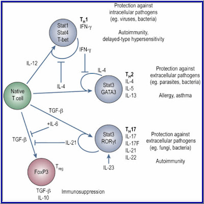
CD4+ T cells regulate immune responses by producing various cytokines upon antigen stimulation. Naive CD4+ T cells have limited cytokine responses and secrete only IL-2 before they differentiate into various effector cell types CD4+ T cells regulate immune responses by producing various cytokines upon antigen stimulation. Each cell type is competing for a limited amount of IL-2 secreted by a small subset of the T helper cells. Naive T cells stimulated with antigen in the presence of IL-4 differentiate into Th2 cells secreting IL-4, IL-5, IL-10, and IL-13, whereas IL-12 induces the differentiation of naive cells into Th1 cells secreting IL-2, IFN-γ, and lymphotoxin.


FROM:
Autoimmunity: IL-21: a new player in Th17-cell differentiation
Elissa K Deenick and Stuart G Tangye
WWW.Nature.com

Ribas and Restifo et al found that if you use Anti-CTLA-4 blockade the native T-cell is pushed towards the Th17 differentiation. This causes less interleukin-2 to be produced by the CD4+ T-cells causing a shortage of IL-2 in the microenvironment for the expanding T-cells thus limiting the differentiation to Tregs. It is well known that in the presence of TGF-beta and IL-2, the Native T-cell differentiates into T Regulatory Cells.

Also the activation and differentiation of Naïve CD8+ T cells require IL-2 provided by activated CD4+ T cells at the initial priming stage within 0–2.5 hours after stimulation. Thers is
critical IL-2 signal from CD4+ cells is mediated through the IL-2R (receptor) of Naïve CD8+ cells. This activation of IL-2 signaling advances the restriction point of the cell cycle, and thereby expedites the entry of antigen-stimulated Naïve CD8+ T-cell into the S phase of the cell cycle.

The cell cycle has four stages:

1. G1 phase when the cell increases in size and gets ready to replicate its DNA.
2. S phase when the cell synthesizes or copies its chromosomes
3. G2 phase in which the cell prepares to divide
4. M phase when mitosis occurs.

If no IL-2 is available to Naïve CD8+ T-cells, they never get a chance to expand and differentiate into Cytotoxic T Lymphocytes (CTLs) Killer T-cells.
Besides promoting cell proliferation, IL-2 stimulation increases the amount of IFNγ and granzyme B produced by CD8+ T cells.

Activation and Differentiation of (CTLs) Cytotoxic T Lymphocytes

Naïve CD8+ T-cells are referred to as CTL-precursors (CTL-Ps), which are incapable of performing any function other then recognizing the class I MHC-antigen complex on the Tumor cells through the (TCR) T Cell Receptor.

For activation, the CTL-P needs at least three signals:

1. Antigen specific signal transmitted by the MHC I peptide/TCR complex for the recognition of the Antigen

2. The Costimulatory signals transmitted by the CD28 receptor and the B7 molecule of the Antigen Presenting Cells (APCs)

3. Cytokine induced signal, IL-2 interaction with the IL-2r (receptor) on the CTL-P leading to activation and differentiation of the CTL-P into effector CTL

The Cytokine IL-2 came from the(CD4+) TH1 cells which means the CTL-P is IL-2 limited and can only be activated by the secreted IL-2 if there is any to be had or by introducing IL-2 through IL-2 therapy. Now you know the reasoning behind using the IL-2 therapy as the second part of the combinatorial Therapy. If there is not enough IL-2 in the host environment, you will only get partial expansion of the CTLs. It may not be enough to eradicate large bulky tumors.

The Interluekin-2 plays another role in this Melanoma Maze. In a study by Janas et al, Il-2 increases the expressions of the perforin and granzyme A, B and C genes in the CD8+ T-cells. This increase expression causes the CD8+ T-cells to mature into Cytoxic T Lymphocytes (CTLs). The exogenous IL-2 is required for the granzyme proteins. CTLs have cytoplasmic granules that contain the proteins perforin and granzymes. A dozen or more perforin molecules insert themselves into the plasma membrane of target cells forming a pore that enables granzymes to enter the cell. Once in the tumor cell, these enzymes are able to breakup (lyse) the cell and destroy it. This is the beginning of the end for the cancer cells. The tumors begin to shrink and the rest is history.

Research suggests that the primary mode of the destruction of the tumors by CTL is by initiating death through the Fas-FasL pathway. Studies have shown that CTLs store Cytotoxic proteins in the form of granules in their cytoplasm.

These proteins belong to two categories:


1. Perforins: involved in pore formation
2. Granzymes: responsible for hydrolysis of the cellular products.

Granzymes breaks down the tumors cells just like your detergent enzymes in your laundry detergent.

Immediately following a CTL contact with the tumor cell, the Golgi sacks load with granules and granzymes which create pores to allow the granzymes to enter and destroy the tumors cells.

So now you know why Anti-CTLA-4 blockage and HD Interluekin-2 go hand and hand. Timing of the addition of IL-2 can make or break the immune response. And without suppressing the Tregs, your chances to mount an immune response may be slim at best. Your Immune System is a well Orchestrated system of events; we just need the Knowledge on how to harvest its potential on eradicating cancer.


The beginning of knowledge is the discovery of something we do not understand.
~Frank Hebert~
A critically acclaimed and commercially successful American science fiction author


Take Care,

Jimmy B
Photobucket

Wednesday, March 17, 2010

The Making of an Immune Response by Combinatorial Therapy Using Anti-CTLA-4 Blockade and Interleukin-2..Melanoma..Jim Breitfeller

The Making of an Immune Response by Combinatorial Therapy Using Anti-CTLA-4 Blockade and Interleukin-2

The paper that I am about to present to you is a culmination of research that was done base on my own experience as a stage IV Melanoma Patient. I wanted to know why my immune system responded and prove to myself and my doctors that it was not a statistical fluke. I dedicate this paper to all my fellow Melanoma Patients that lost their battle with the Beast, especially Bob Luker who fought so bravely but was unable to obtain Anti-CTLA-4 blockade due to the shortage proclaimed by Bristol Myer Squibb which halted compassionate drug use on 9-12-2008.

Most Melanoma tumors are accepted by the host’s immune system and progress even when they contain potentially antigenic proteins. This may be due to the tumor secreting immunosuppressive Cytokines like, TGF-Beta, IL-10, IL-4 and IL-6. TGF-β inhibits the proliferation and functional differentiation of T lymphocytes. TGF- Beta accelerates the expression of CTLA-4 by stimulated CD4+CD25– T cells. TGF- Beta requires CTLA-4 early after T Cell Activation to induce FoxP3 and generates adaptive CD4+CD25+ (Treg) Regulatory Cells. The tumor cells secrete TGF-Beta.

The Making of an Immune Response by Combinatorial Therapy Using Anti-CTLA-4 Blockade and Interleukin-2


Take Care,

Jimmy B
Photobucket

Tuesday, March 9, 2010

Challenges and Opportunities Ahead..Melanoma Therapy ..Jim Breitfeller

"A dynamic picture is emerging in which the immune system is far from quiescent in cancer, and tumor cells elaborate a host of countermeasures to evade immune destruction. In essence, these are two systems which co-evolve over time. Clinical outcome is ultimately a balance between the proliferation of tumor cells and the immune response which attempts to control these cells. Various treatments may perturb these two systems in different ways - in good clinical outcomes, the immune response ultimately dominates, while in poor outcomes, tumor cells dominate.

It is becoming clear that the challenge in tumor immunotherapy lies not only in lack of tumor recognition, but also ineffectivity of the immune response that develops. While efforts to elicit tumor-specific T cells directly in patients via vaccination may overcome potential mechanisms of immune evasion by tumors cells, current immunotherapeutic strategies do not address their potential immunomodulatory mechanisms.

Understanding the molecular mechanisms which underlie T cell dysfunction in cancer will be important to devise novel adjunct strategies to enhance or modulate T cell responses after tumor-specific T cells have been elicited. It will likely take a multi-faceted approach acting at several steps along the T cell activation and effector activity pathway to finally achieve success in cancer immunotherapy."


The Laboratory of Peter P. Lee, MD at Stanford University

This multi-faceted approach acting at several steps along the T cell activation and effector activity pathway is what I call Combinatorial Therapy.

We first start at obtaining the Antigen to be presented on the Antigen Presenting Cell. Then once we think we got it, we need to proceed to the Activation step. Ipilimumab (Anti-CTLA-4 Blockade) helps to keep the activation in the on position. It also has a dual role, in keep the Treg's surpressive function out of play in the Tumor 's microenvirioment.
Interleukin -2 is the final step to complete response. It is the growth factor for the CTL's, which help maintain functionality and survival of the Cytotoxic T Lymphocytes (CTL's).


Take Care,

Jimmy B
Photobucket

Thursday, February 25, 2010

A note To Dr. Keith Flaherty.. Combinatorial Therapy..Melanoma..Jim Breitfeller..2-25-2010

"One year, Dr. Flaherty thought, when he heard the news. Certainly no triumph. But it was something. Something to be built on.

Novartis and Bristol-Myers had agreed to schedule teleconferences for later in the month to talk about combination trials. He checked the dates on his electronic calendar. A meeting with Pfizer was also pending."


Dr. Flaherty,

As early as May of 2009 I had come to the conclusion that the drug companies must work togther to come up with a stabilization/cure.

As a fellow researcher and Blogger, What can I do to help convince the pharma this it is in their best interest to work together. It would be a win, win for all, including the Patients like myself.

Keith, Please let me know what I can do to Help?

Best regards


Jim Breitfeller


Friday, May 8, 2009

There is Enough Room for Novartis, Bristol Meyer Squibb and Pfizer for each to take part in the Melanoma Space Melanoma.. Jim Breitfeller

There is Enough Room for Novartis, Bristol Meyer Squibb and Pfizer for each to take part in the Melanoma Space.

With ASCO Annual meeting coming up at the end of May, I believe that there will be some excitement around monoclonal antibodies. In particular, anti-CTLA-4 blockage treatment will be under the spotlight. What will the overall survival rates look like? Well, speaking from my experience, anti-CTLA-4 has prolonged my life for over 27 months where as a stage IV Patient, I was given 6 to 9 months. This could not have happen with out the extra help from Interleukin -2.

See, once the CD4+ T-cells activated, they need to grow and cross-prime the CD8+ T-cells. While activated T-cells secretes IL-2 at a certain concentration to help promote proliferation of the CD4+ T-cells. If there is too much IL-2 at the beginning of the immune response, I believe that the ratio of CD4/CD8/CD3 would be altered causing
the Tregs (CD4+ CD25 fox P3) to gain the upper hand of suppression of the immune response. A subset of CD4 + cells called CD4 + CD25 + regulatory T (Treg) cells that expresses Forkhead box P3 (Fox P3).

With just adding Anti-CTLA-4 first, It has been reported that it suppresses the T regs and pushes the balance towards an immune response. Once the immune response is in progress, the CD4+ T-cell is needed to co-stimulate the CD8+ T-cell. A source of these cofactors for effective CD8+ T-cell stimulation can be provided by CD4+ T cells that release critical amounts of IL-2.

Once the CD8+ T-cell is activated, and is cultured in the presence of Interleukin 2, it results in the development of effector cells which are cytotoxic to tumor cells.

This is why we the patients need the Pharmacitical companies to work together. Each therapy will not work alone as a single agent. The response rate are between 10 and 22 percent for each therapy. If you do a sequential treatment with the proper dosage and timing, you will see a synergistic outcome.


“There are three parts of the equation in a clinical trial. There’s who has control, who gets the reward, and who takes the risk. Patients take all the risk, they have no control, and they get no reward. Patients ought to be the ones driving the process and get the reward out of it and have the control, since they are the ones that take the risk.”

~Greg Simons~


If we are taking all the risk, shouldn’t we have a say in our Destiny?

“We need to all work together for the common good of the Melanoma Patients”

“We need to take greed out of the equation and just do what is right for humanity”


Melanoma and The Magic Bullet (Monoclonal Antibodies)


Take Care,

Jimmy B
Photobucket

Thursday, February 4, 2010

The Orchestration of an Immune Response Unrehearsed! Melanoma..Jim Breitfeller

February 4 is World Cancer Day—a global day of awareness created by the International Union Against Cancer. With cancer set to become the #1 killer in the world this year, the day brings us together to highlight the growing personal and economic impact of the disease. On this day it’s critical that each organization—and each individual—share responsibility for sending a powerful message about cancer prevention.


Melanoma And the Magic Bullet (monoclonal Antibodies


Take Care,

Jimmy B
Photobucket

Greetings to One and All

This Blog is dedicated My Brother Kenny B. who passed away in the late 1970's with Cancer before the Internet.

It was he, who showed me How to live and give back. He was wise beyond his years.


Kenny B


Jimmy and Dee

Carepage: Jimmybreitfeller
Jimmy Breitfeller


My Profile as of 2009

My photo
Last July (2005)I was riding my bicycle to work at the Eastman Kodak Research Labs about 3 miles from home. I was wearing a knapsack to carry my things to and from the labs. I started noticing an ache on my back. So I decide to go to the dermatologist. To make the long story short, it was cancer. I knew from my research that I would be needing adjuvant therapy. So I started communicating with Sloan Kettering, University of Pittsburgh Cancer Center, and a couple of others including the Wilmot Cancer Center at Strong. I realized that by telling my story, I might help someone else out there in a similar situation. So to all who are linked by diagnosis or by relation to someone with melanoma, I wish you well. Stay positive, read as much as you can (information helps to eliminate the fear associated with the unknown), and live for today, as no one can predict what tomorrow may bring. Jimmy B. posted 12/15/08

Disclaimer

The information contained within this Blog is not meant to replace the examination or advice of your Oncologist or Medical Team. The educational material that is covered here or Linked to, does not cover every detail of each disorder discussed.

Only your physician/Oncologist can make medical decisions and treatment plans that are appropriate for you. But, An Educated Consumer is a Smart consumer.

As Dr. Casey Culberson Said:

"The BEST melanoma patient is an ACTIVE PARTICIPANT in his or her treatment
(not a PASSIVE RECIPIENT)"

Melanoma and the “Magic Bullet” (Monoclonal Antibodies)

Just to let you know I posted the first draft of the Melanoma and the “Magic Bullet” (Monoclonal Antibodies). on Melanoma Missionary In the Shared File Section. you can download it for 19.95 (Only kidding) it is Free for the taking.


It is 33 pages long and may help you in your quest for the Yellow Brick Broad. Just to let you know it is only the first draft. Revisions are sure to come. I wanted to get it to the people that need it the most, the Melanoma Patients.

Preview:

So, where does Interluekin-2 (IL-2) come into play? According to Byung-Scok et al and recent reports, IL-2 is not needed for developmental CD4+ CD25+ Treg cells in the thymus but does play an important role in the maintenance and function in the peripheral.18 Peripheral is defines as secondary system outside the bone marrow and thymus. It entails the site of antigen, immune system interaction. IL-2 is required for the peripheral generation of Tregs based Abbas’s and colleagues research.19

IL-2 prevents the spontaneous apoptosis of the CD4+ CD25+ Treg cells. It has been reported that patients with multiple advance-stage tumors have elevated levels of Tregs within the tumor microenviroment.20 Interluekin-2 is the survival factor for CD4+ CD25+ Treg cells.21 If the addition of IL-2 is on or before the maximum propagation of the CD4+ T cells, the Tregs population can increase 5-fold in a 96 hour period based on certain growth mediums.

By controlling the addition of the endogenous IL-2, one has a knob to turn and can lead to the control of the expansion of the Tregs. When you combined this control with the anti-CTLA-4 blockage, you can shift the balance of the immune response.

Now here is the catch. The maintenance and function of the CD8+ T-cells require CD4+ cells which secrete IL-2. So we don’t want to deplete the CD4+ cells, we want to control the expansion of the Tregs which are a subset of the CD4+ cells. It has been postulated by some researchers that the Anti-CTLA-4 blockage also suppresses the Treg function in a different mechanism. By using IL-2 as the rate limiting factor, we can suppress the CD4+ CD25+ Treg cell expansion by controlling the concentration and timing of the Inerluekin-2 at the tumor microenvironment.


The Interluekin-2 plays another role in this Melanoma Maze. In a study by Janas et al, Il-2 increases the expressions of the perforin and granzyme A, B and C genes in the CD8+ T-cells. This increase expression causes the CD8+ T-cells to mature into Cytoxic T Lymphocytes (CTLs). The exogenous IL-2 is required for the granzyme proteins. As stated previously, CTLs have cytoplasmic granules that contain the proteins perforin and granzymes. A dozen or more perforin molecules insert themselves into the plasma membrane of target cells forming a pore that enables granzymes to enter the cell. Once in the tumor cell, these enzymes are able to breakup (lyse) the cell and destroy it. This is the beginning of the end for the cancer cells. The tumors begin to shrink and the rest is history,


On the other hand, prolong therapy with Il-2 can result in causing apoptotic death of the tumor- specific CD8+ T-cells.23

Clearly in a clinical setting, timing, dose, and exposure to these drugs play a major roll in the immunotherapy, and can have dramatic effects on the outcome.

All it takes is that one magic bullet to start the immune reaction..

https://app.box.com/shared/kjgr6dkztj

Melanoma And The Magic Bullet (Monoclonal Antibodies)

Public Service Announcement

A call for Melanoma Patients by Dr. Steven A Rosenberg

"We continue to see a high rate of clinical responses in our cell transfer immunotherapy treatments for patients with metastatic melanoma", Dr. Rosenberg said.

"We are actively seeking patients for these trials and any note of that on a patient-directed web site would be appreciated."

If you would like to apply for his trials, here is the website and information.

Dr. Rosenberg's information


Dr. Rosenberg's Clinical Trials


For the Warriors


The Melanoma Research Alliance has partnered with Bruce Springsteen, the E Street Band, and the Federici family to alleviate suffering and death from melanoma. Please view Bruce Springsteen’s public service announcement inspired by Danny Federici. Danny was the E Street Band’s organist and keyboard player. He died on April 17, 2008 at Memorial Sloan-Kettering Cancer Center in New York City after a three year battle with melanoma.


http://www.melanomaresearchalliance.org/news/PSA/

Source Fastcures blog


Join the Relay for Life!!!

Photobucket

Dear Family and Friends,

I’ve decided to take a stand and fight back against cancer by participating in the American Cancer Society Relay For Life® event right here in my community! Please support me in this important cause by making a secure, tax-deductible donation online using the link below.

To donate on line now, click here to visit my personal page.
Jimmy B AKA Melanoma_Missionary

Relay For Life® is a life-changing event that brings together more than 3.5 million people worldwide to:

CELEBRATE the lives of those who have battled cancer. The strength of survivors inspires others to continue to fight.

REMEMBER loved ones lost to the disease. At Relay, people who have walked alongside people battling cancer can grieve and find healing.

FIGHT BACK. We Relay because we have been touched by cancer and desperately want to put an end to the disease.

Whatever you can give will help - it all adds up! I greatly appreciate your support and will keep you posted on my progress.

Keep the Fire Burning!!!

Photobucket

Sincerely,

Jimmy Breitfeller
Turn off Music before you "Click to Play"
Signs of Melanoma Carcinoma Skin Cancer

How Skin Cancer Develops by "About.com : Dermatology"

Call for Patients with Unresectable Liver Metastases Due to Melanoma


Delcath Systems Granted Orphan-Drug Designations for Cutaneous and Ocular Melanoma


Delcath is actively enrolling patients in a Phase III clinical trial testing its proprietary drug delivery system, known as Percutaneous Hepatic Perfusion (“PHP”), with melphalan for the treatment of ocular and cutaneous melanoma metastatic to the liver.

This NCI-led trial is enrolling patients at leading cancer centers throughout the United States. Commenting on these orphan-drug designations, Richard L. Taney, President and CEO of Delcath, stated, “These favorable designations are important steps in our efforts to secure Delcath’s commercial position upon conclusion of our pivotal Phase III trial for metastatic melanoma. We remain steadfast in our commitment to become the leader in the regional treatment of liver cancers and we continue to enroll patients in this study, and advance our technology and the promise that it offers to patients with these deadly forms of melanoma and other cancers of the liver, all with limited treatment options.”

Orphan drug designation, when granted by the FDA’s Office of Orphan Products Development, allows for up to seven years of market exclusivity upon FDA approval, as well as clinical study incentives, study design assistance, waivers of certain FDA user fees, and potential tax credits.


Current Trial Centers


Phase I Study of Hepatic Arterial Melphalan Infusion and Hepatic Venous Hemofiltration Using
Percutaneously Placed Catheters in Patients With Unresectable Hepatic Malignancies


James F. Pingpank, Jr., MD, FACS
Associate Professor of Surgery
Division of Surgical Oncology
Suite 406, UPMC Cancer Pavillion
5150 Centre Avenue
Pittsburgh, PA 15232
412-692-2852 (Office)
412-692-2520 (Fax)
PingpankJF@UPMC.edu


Blog Archive

Call For Melanoma Patients!!!!

Call For Melanoma Patients!!!!

Dr. Rosenberg Has a New Clinical Trial.

Our latest treatment has a 72% objective response rate with 36% complete responses.

We are currently recruiting patients for our latest trial.

Is there some way to post this “Call for Patients” on the web site?

Steve Rosenberg

Dr. Rosenberg's Clinical Trials


(For a copy of the research paper.. see My Shared files)

The news headlines shown above for Melanoma / Skin Cancer are provided courtesy of Medical News Today.